
Introduction
Medical imaging plays a crucial role in diagnosing various conditions, and Colour Doppler ultrasound is one of the most effective tools for assessing blood flow. Unlike traditional grayscale ultrasounds, Colour Doppler provides real-time visuals of how blood moves through arteries and veins.
This technology is widely used in cardiovascular, obstetrics, and vascular health to detect blockages, clots, and abnormal blood flow patterns. The color mapping technique used in this scan helps doctors visualize the direction and speed of blood movement, making it an essential diagnostic tool.
If you are looking for a color doppler test in Mumbai, consulting a trusted diagnostic center like Midas Care Clinic can ensure accurate results.
Understanding Colour Doppler
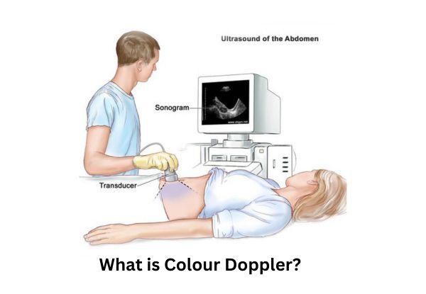
What is Colour Doppler?
Colour Doppler ultrasound is an advanced imaging technique that helps monitor blood flow in real-time. It differs from traditional grayscale ultrasound by incorporating color-coded images, allowing medical professionals to evaluate circulation efficiency, blockages, and abnormalities.
Unlike conventional B-mode ultrasound, which only displays static images, Colour Doppler adds motion analysis by showing blood flow speed and direction using red and blue color signals.
How Does Colour Doppler Work?
The Doppler effect is the principle behind this imaging technique. It measures how sound waves bounce off moving blood cells and return to the ultrasound probe. This shift in frequency helps determine the direction and velocity of blood flow.
In a color doppler sonography, different colors indicate specific movements:
- Red signals show blood flowing toward the probe.
- Blue signals indicate blood moving away from the probe.
This color-coded representation helps doctors detect irregular blood circulation, ensuring early detection of vascular conditions.
Types of Doppler Ultrasound
Colour Doppler Ultrasound
This technique provides detailed visuals of blood flow patterns and is commonly used in cardiovascular and fetal imaging.
Power Doppler Ultrasound
A more sensitive version of Colour Doppler, it helps detect low-speed blood flow, particularly useful for evaluating small blood vessels and inflamed tissues.
Spectral Doppler Ultrasound
Unlike color mapping, Spectral Doppler produces a graphical representation of blood velocity over time, offering quantitative data about circulation.
Duplex & Triplex Doppler Ultrasound
These methods combine grayscale ultrasound, color mapping, and spectral waveforms to provide comprehensive vascular assessments.
Uses & Applications of Colour Doppler
Medical Applications
Colour Doppler is instrumental in diagnosing various vascular conditions such as:
- Deep vein thrombosis (DVT): Detecting blood clots in veins.
- Atherosclerosis: Identifying artery narrowing due to plaque buildup.
- Varicose veins: Assessing valve dysfunction in leg veins.
- Carotid artery disease: Checking for obstructions that may lead to strokes.
Obstetrics & Gynecology Applications
This imaging method is crucial in pregnancy monitoring, helping assess:
- Fetal blood flow to ensure proper oxygen and nutrient supply.
- Placental health, detecting complications like insufficient blood circulation.
Cardiovascular & Renal Applications
Doctors use Colour Doppler for detecting:
- Heart valve abnormalities affecting blood circulation.
- Renal artery stenosis, a condition that affects kidney function.
For individuals seeking accurate diagnostic imaging, consulting a specialist like Dr. Chandrakant ensures expert evaluation.
Procedure: What to Expect During a Colour Doppler Scan?
Preparation Before the Test
- Most scans do not require fasting, but abdominal tests may need a few hours of fasting.
- Loose, comfortable clothing is recommended for easy access to the scanned area.
How is the Procedure Performed?
- A technician applies gel to the skin and uses an ultrasound probe to capture images.
- The scan typically lasts 15–45 minutes, depending on the area being examined.
Post-Test Considerations
- Results are analyzed to detect abnormalities.
- A doctor may recommend further testing based on findings.
Patients looking for a reliable diagnostic service can visit the Best diagnostic center in Vasai for expert consultation.
Benefits & Risks of Colour Doppler
Benefits
- Non-invasive & painless, No incisions or needles required.
- Real-time blood flow analysis for accurate diagnosis.
- No radiation exposure, making it safe for repeated use.
Risks & Limitations
- Limited penetration in obese patients, reducing image clarity.
- Results depend on the technician’s expertise, requiring skilled professionals for interpretation.
Frequently Asked Questions
Q1: Is Colour Doppler the same as a regular ultrasound?
Ans: No. A color doppler sonography provides real-time blood flow visualization, unlike grayscale ultrasound, which only captures static images.
Q2: How long does a Colour Doppler test take?
Ans: A color doppler test in Mumbai typically lasts 15–45 minutes, depending on the area being examined.
Q3: Is Colour Doppler painful?
Ans: No, it is a non-invasive and painless procedure that involves only mild pressure from the ultrasound probe.
Q4: Can a Colour Doppler detect heart problems?
Ans: Yes. It is used to diagnose heart valve defects and detect abnormal blood circulation in arteries.
Q5: Do I need any special preparation for a Colour Doppler test?
Ans:
- For most scans, no preparation is needed.
- Abdominal scans may require fasting to improve imaging clarity.
Conclusion
Colour Doppler ultrasound is a powerful diagnostic tool that plays a critical role in detecting vascular diseases, pregnancy complications, and cardiovascular issues.
Its non-invasive nature and real-time imaging capabilities make it a preferred choice for both doctors and patients. If you are experiencing circulatory problems or require a routine blood flow assessment, a color doppler test in Mumbai at Midas Care Clinic can provide accurate and reliable results.
For expert guidance, consider consulting Dr. Chandrakant, a specialist in vascular imaging.